

Outline
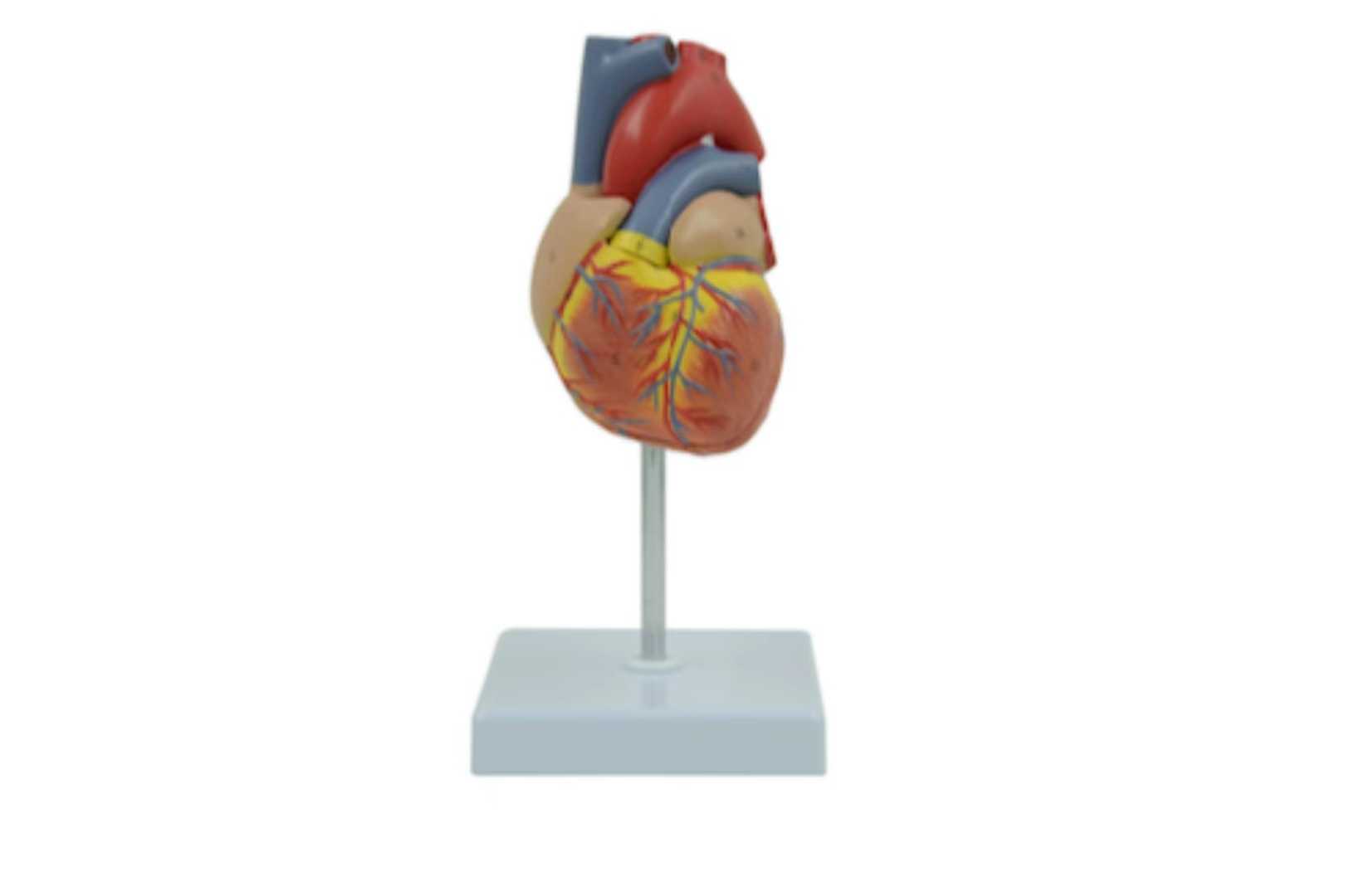
2-Part Life-Size Human Heart Model is an anatomically correct representation of the human heart. The deluxe model features a removable heart wall to display internal structures, exposing the hollow cavity of the heart and the anatomical structures that border and intersect between the muscular heart walls. The model’s removable heart wall contains raised and colored vasculature against the muscle.
The model includes a removable desktop base stand, making it perfectly compact for display and study anywhere. This high-quality heart anatomy model was designed by medical professionals and is perfect for medical students, educators, and professionals.
Skills Gained
· Familiar with heart anatomy
Features
Deluxe Life-Size Heart Model
The 2-Part Life-Size Human Heart Model is a deluxe human heart replica. The heart model features raised veins and arteries and textured muscle striations to provide a tactile study of the organ. Once detached from the base stand, the model can be viewed to display all 5 surfaces of the heart: anterior, posterior, diaphragmatic, right pulmonary, and left pulmonary.
Major external arteries, veins, and trunks are represented on the model, including the superior vena cava, ascending aorta, aortic arch, pulmonary veins and arteries, and the brachiocephalic trunk. These anatomically correct external structures are perfect for studying the blood flow to and from the heart.
2-Part Heart
The 2-part heart is made up of the removable front heart wall and the main body of the heart model. This detachable part of the model displays the external surface of the left and right ventricles, the left and right auricle, the great cardiac vein, the anterior interventricular artery, right coronary artery, and cardiac muscle. These anatomical features are displayed in correct positioning and layers. The front heart wall is held together by discreetly hidden magnets for an uninterrupted view of the model.
Once the front heart wall is removed, the hollow cavity of the heart is exposed. In between these spaces are the major structures found in the heart’s internal anatomy. These structures include the tricuspid valve, bicuspid valve, and the interior compartments of the left and right ventricle and atrium. Additional detail includes the interventricular septum, which is the dividing wall between the chambers.
Previous: Urinary System
Stock code :833047
Address:2nd & 3rd Floor, West 6th Building, 18 West HaiTai Road, Tianjin, China
Postcode:300384
Phone:4006-355-510
+86-22-83711066
Fax:+86-22-83711065
Email:info@tellyes.com

 Anatomy